Data Acquisition Technology Permits Fast Optical Recording for Studies of Causes of Sudden Cardiac Death
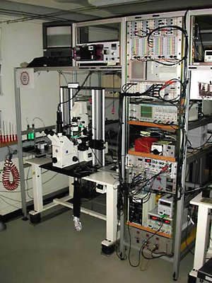
Figure 1, photograph of measurement setup
The optical recording setup is built around an inverted microscope equipped for epifluorescence. The emitted fluorescence of the preparation is projected onto the input window of an array of fiber optic cables attached to the bottom port of the microscope. Individual fibers are connected to individual photodetectors and, after analog signal conditioning, the signals are routed to the multiplexers of the DAPs, which are mounted in the rack above the PC.
By using self-contained data acquisition boards instead of conventional analog-to-digital (A/D) converters, Dr. Stephan Rohr of the University of Bern was able to develop a unique optical recording system for studying cellular mechanisms underlying sudden cardiac death. Rohr's system advances this field of research beyond studies of whole heart tissue by using fiber optics to study impulse propagation at the cellular level. An A/D converter with an onboard microprocessor is a critical element of the system because it permits the gathering of data from over 200 photodetectors in parallel at very high speed. Rohr has equipped his system with three of these devices, called data acquisition processors (DAPs), which perform data conversion simultaneously with data collection and write the results to a hard disk. "Previous A/D cards had to interact with the computer's operating system, which tended to disrupt measurements," says Rohr, a researcher in the Swiss university's department of physiology. "Because DAPs are self-contained, they perform the data conversion very reliably and aren't affected by what the host computer is doing."
Dr. Rohr's work, supported by the Swiss National Science Foundation, is based on the previously established premise that slow conduction of electrical impulses through the heart is one of the key mechanisms in the generation of cardiac arrhythmia. Knowing that alterations in the cellular architecture of cardiac tissue contribute to slow conduction, Rohr has undertaken a systematic investigation of the relationship between altered cellular tissue architecture and impulse conduction. "In normal structured heart tissue, impulse propagation from cell to cell is fast and uniform, ensuring the normal pumping function of the heart," Rohr explains. "In hypertrophied or older hearts, fibrous tissue can be inserted between the layers of cardiac cells. This structure is not uniform anymore and the electrical impulse doesn't travel uniformly. It takes unpredictable twists and turns, which can lead to generation of arrhythmias."
Insights into Arrythmia
By using [DAP boards], Dr. Stephan Rohr of the University of Bern was able to develop a unique optical recording system for studying cellular mechanisms underlying sudden cardiac death. Rohr's system advances this field of research beyond studies of whole heart tissue by using fiber optics to study impulse propagation at the cellular level.
“Previous A/D cards had to interact with the computer's operating system, which tended to disrupt measurements. Because DAPs are self-contained, they perform the data conversion very reliably and aren't affected by what the host computer is doing.”
Dr Stephan Rohr, University of Bern
Previous technical limitations
Before Rohr could study impulse propagation at the cellular level, he had to overcome a number of technological limitations that had previously restricted researchers to working with whole heart tissue only. The assessment of electrical activity at the cellular/subcellular level requires the use of voltage-sensitive fluorescent dyes. Changes in fluorescence are linearly related to changes in voltage, so one of the requirements for the research is to be able to detect changes in fluorescence. This is normally done with photodetectors. One limitation Rohr ran into in trying to work at the cellular level related to the type of photodetector used. The accepted method at the time used CCD cameras to record macroscopic changes in fluorescence. This worked for whole heart measurements because the changes in fluorescence were relatively large and propagation times were long enough to permit the determination of activation patterns at the slow (60 Hz) frame rates of these devices. The monolayer of cells that Rohr wanted to work with produced minimal changes in florescence. And even the fastest cameras could not capture impulse propagation at the cellular level.
This problem was solved by replacing a CCD camera with photodiodes connected to a freely selectable subset of 379 plastic-coated, 1-mm fiber optic cables. This fiber optic recording technique delivers sufficiently high spatial and temporal resolution to follow impulse propagation at the cellular and even subcellular level. In Rohr's system, photocurrents captured by the photodiodes are converted to voltages by first stage amplifiers. After a second amplifier subtracts background fluorescence, the signal is filtered and amplified again. At this point the signal is transferred to an A/D converter in a PC.
|
Figure 1, photograph of measurement setup |
Rohr had another technical hurdle to overcome with A/D conversion. His first system was equipped with two A/D cards that together allowed him to collect data from 80 channels. His goal, however, was to collect data from over 200 photodiodes at once. In addition to limiting the number of channels, the previous cards also limited how much data could be collected in any given session. This is because they moved data back and forth between the PC's RAM and had no way to off-load data to a storage device. When RAM was filled the experiment had to cease.
Another drawback to those early A/D boards was that they had to interact with the PC's operating system. This was a problem for Rohr for two reasons. First, when the operating system changed, he had to rewrite his test routine. "I never knew when Microsoft would change the operating system, and when they did it took a lot of time until the system was running properly again," he explains. The other problem was the fact that Windows is not a real-time operating system. Running the A/D conversion under Windows made the system vulnerable to situations where Windows was occupied with other tasks, possibly interrupting the data flow. "Operating systems like Windows use up many cycles on the PC platform, and when they take control of the CPU they hold onto it for a long time in data acquisition terms," says Rohr. "To convert the data in real-time, computing power has to be available whenever it is needed."
Bypassing Windows
Rohr needed to be able to perform A/D conversion without involving Windows or the PC's CPU. He wanted to be able to add channels as necessary without the need to completely reconfigure his system. And he wanted the ability to transfer the digital data produced by A/D conversion to the PC's hard disk so the experiment could proceed for longer lengths of time. These requirements led him to investigate other options for A/D conversion. His research led him to the DAP board from Microstar Laboratories, Bellevue, Washington, which met his requirements.
The unique capability offered by DAPs is an onboard microprocessor that runs a multitasking, real-time operating system optimized for high-performance real-time data acquisition and control applications. The intelligence on the DAP board extends the power of the Windows user interface by executing all processor-intensive routines in real time so that there's never any danger of losing data regardless of how many computer cycles are dedicated to the foreground application or even if Windows crashes. The data acquisition processor continues to run totally independent of the central processor.
Rohr configured his system with three DAPs that run in parallel. A small amount of programming was required to send configuration information to the DAPs -- to indicate the sampling rate, for example, and to set up the high-speed channel from the DAP to the PC for acquired data -- and then to transfer the data to the computer's hard disk. The programming was done using Interactive Data Language (IDL) from Research Systems, Boulder, Colorado. IDL programming also controls the experiment and analyzes the data. Rohr's system uses three DAPs simultaneously to support data collection from over 200 channels. The DAP model he chose includes multiple A/D converters, giving the boards the capability of handling the 20 kHz sampling rate from each of the channels -- a combined data rate of over five MHz -- in real time. If Rohr wants to expand the system in the future with more channels, he can do so simply by adding additional DAPs.
Reconfiguring the optical recording system with DAPs solved the problems Rohr was having with traditional A/D boards, allowing him to collect data from many more channels and thus to visualize electrical activity in a bigger area of heart cell cultures. It also enables him to record for much longer times than when RAM was the limiting factor. The new optical recording system is helping Rohr gain new insights about how cell structure affects impulse propagation. One of his findings, which he describes as "paradoxical," was published in the journal, Science.
In that article, Rohr acknowledged that disturbances in impulse propagation can be caused by structural discontinuities such as "surviving tissue strands connecting islands of intact myocardial tissue in infarct scars." He wrote: "In . . . structural discontinuities, a small current source is connected to a large current load, resulting in a current-to-load mismatch that causes slowing of conduction or conduction block. Both electrical uncoupling and the presence of discontinuous tissue structures are known to be involved in the generation of life-threatening cardiac arrhythmias and it is generally assumed that they act synergistically in the depression of conduction." His findings indicated, however, that "uniform electrical uncoupling occurring in discontinuous tissue structures improves conduction."
These sorts of findings would not be possible without the unique optical recording system. The system is proving to be very valuable in studying the causes of sudden cardiac death, the leading killer in Western society. By using DAPs to overcome the limitations of conventional A/D equipment, Rohr was able to design a system capable of handling the large amount of data necessary to get such a close a look at the heart.
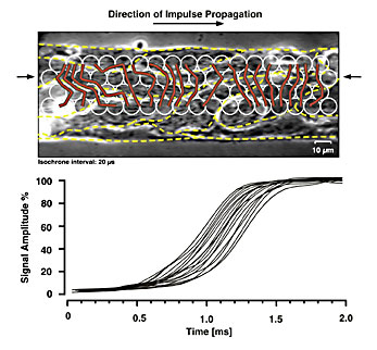
Figure 2, Subcellular assessment of impulse propagation in cardiac tissue |
Browse other customer applications.